
Basic information of the patient
- Gender: Male Age: 33
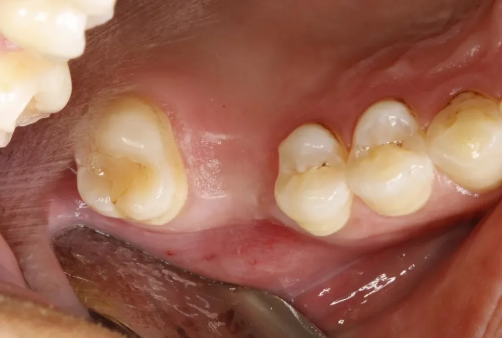
- Chief Complaint: Missing right upper posterior tooth for more than 8 months.
- Present illness: The right upper posterior tooth was extracted from another hospital due to caries over 8 months ago. The patient denied any pain or discomfort. Now, the patient feels that it affects chewing and has come to the hospital for restoration.
- Past History: History of tooth extraction at an outside hospital.
- Clinical Diagnosis: (16) Maxillary dentition defect
Preoperative diagnosis of the patient – Intraoral photograph
Preoperative diagnosis of the patient – Intraoral photograph-CBCT
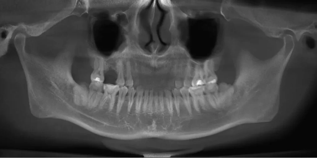
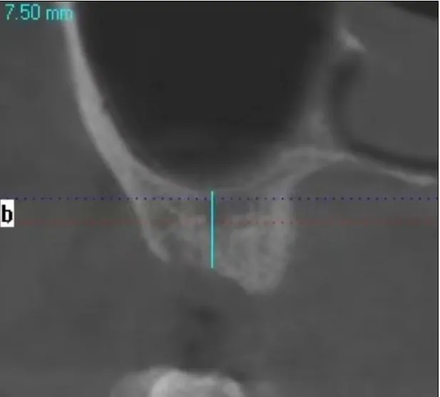
This image intuitively demonstrates the condition of insufficient bone height in the posterior maxillary region (with a residual alveolar bone height of 7.5 mm), representing a typical case that requires maxillary sinus floor elevation prior to implant placement. We selected Rosemond RevoMedx Natural Bovine Bone Graft Material for the sinus elevation procedure, with the goal of providing sufficient osseointegration space and primary stability for the dental implant.
Treatment plan
- Step 1:One-stage simultaneous maxillary sinus floor elevation and implant surgery
- Step 2:Second-stage implant surgery
- Step 3:Implant impression
- Step 4:Implant prosthesis delivery
Preoperative Preparation
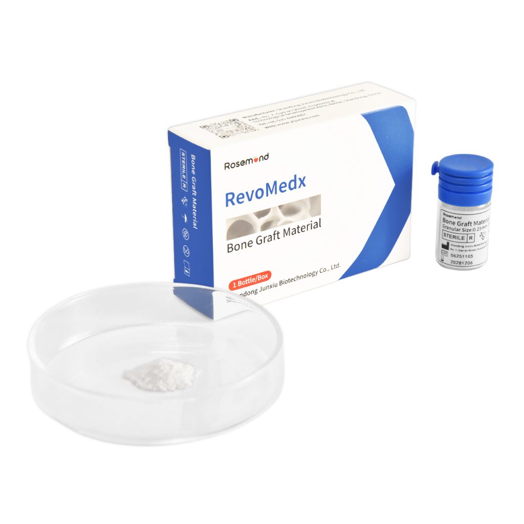
We selected Rosemond RevoMedx Natural Bovine Bone Graft Material for the sinus elevation procedure.
Intraoperative Photograph – Tooth #16
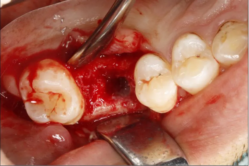
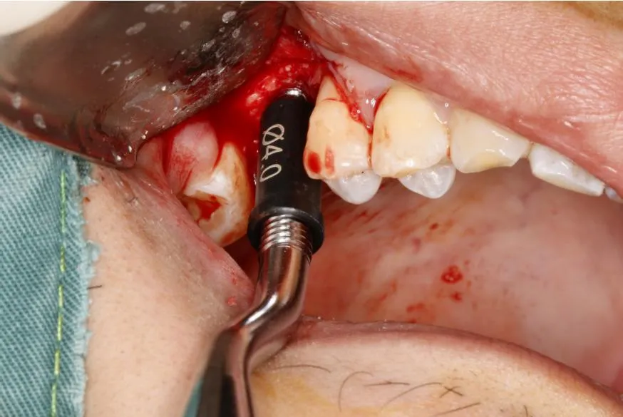
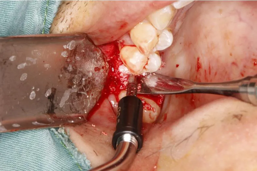
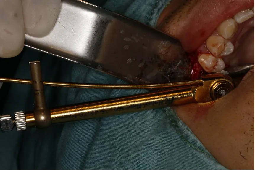
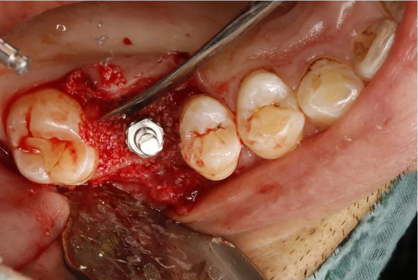
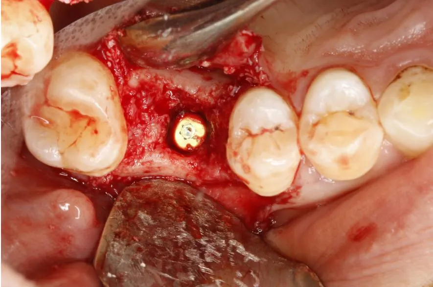
From left to right:
- Exposure of the surgical site and evaluation of bone defect
- Elevation of the maxillary sinus floor membrane
- Placement of bone graft material
- Compaction and contouring of bone graft
- Placement and seating of the dental implant
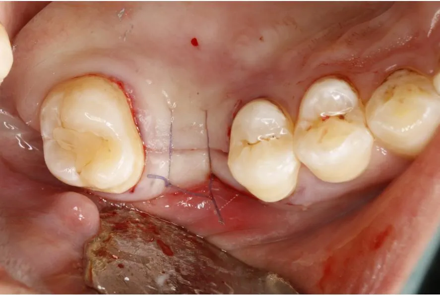
- Wound coverage and closure
Postoperative photo – Tooth position 16
Postoperative CBCT examination – Tooth position 16
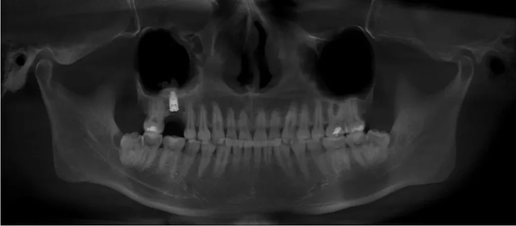
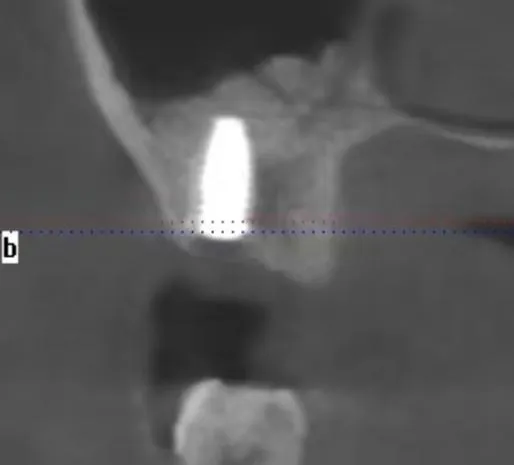
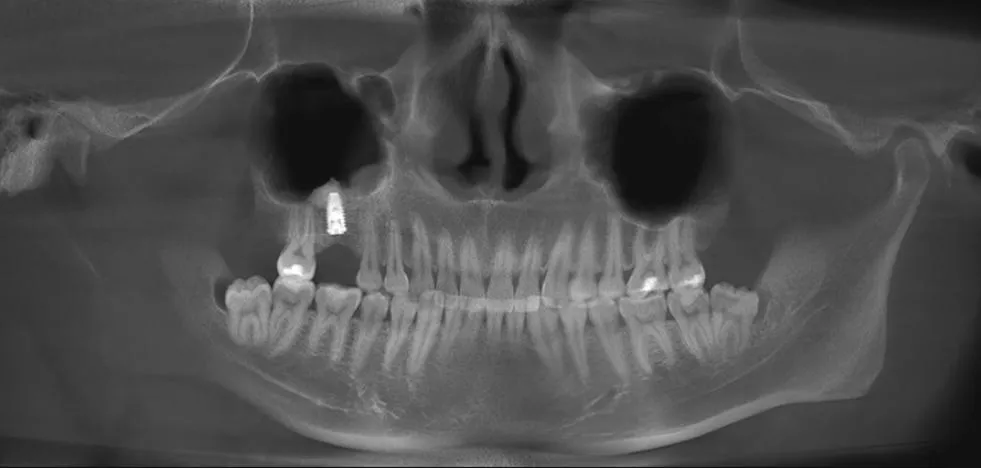
Left (Panoramic View):The postoperative CBCT panoramic view clearly shows the successfully placed dental implant at tooth #16 (maxillary first molar). The implant is in an ideal position, with homogeneous radiopacity of the surrounding bone graft material and intact surgical site anatomy, no implant displacement or abnormal radiopacity is observed.
Middle (Sagittal View):The sagittal cross-sectional image demonstrates the anatomical relationship between the implant and the maxillary sinus floor. The maxillary sinus membrane is intact and elevated, and the bone graft material is adequately filled in the sinus floor space, providing stable bony support for the implant.
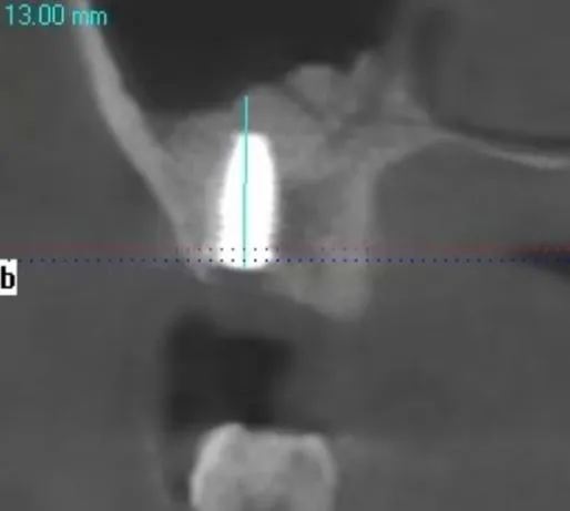
Right (Sagittal Measurement View):The sagittal measurement shows an effective bone height of 13.00 mm around the implant, a significant increase from the preoperative 7.50 mm. This confirms that the maxillary sinus floor elevation procedure successfully augmented the bone volume, ensuring sufficient osseointegration space and primary stability for the implant.
Postoperative CBCT examination at 6 months -Tooth position 16
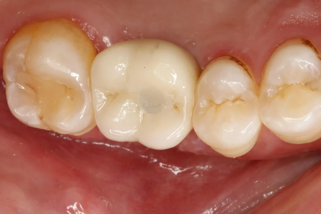
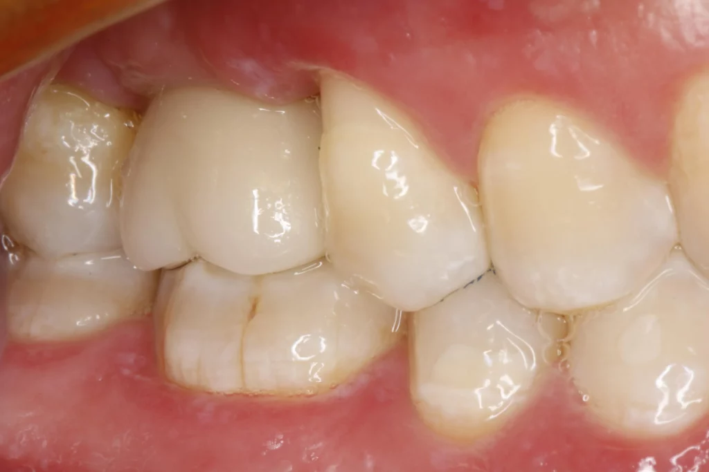
Post-insertion photo – Tooth position 16
Post-insertion CBCT examination – Tooth position 16
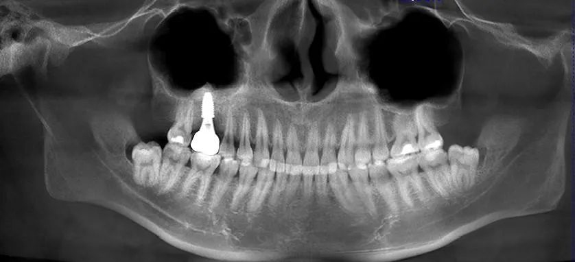
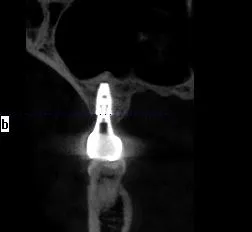
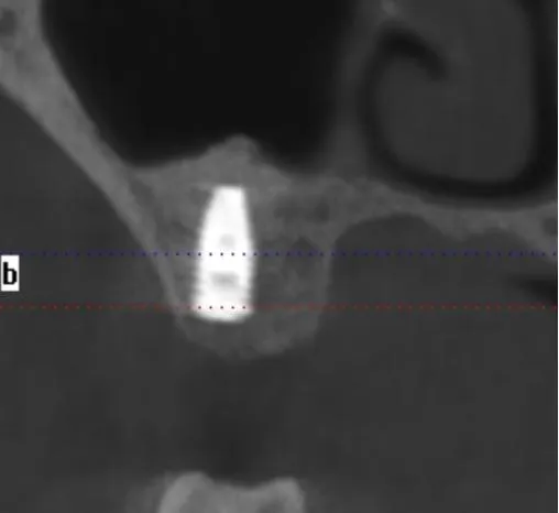
This post-restoration CBCT image clearly demonstrates the final outcome of implant rehabilitation for tooth #16 (maxillary first molar):
- The implant is precisely positioned with stable osseointegration, and the suprastructure is well-seated, achieving optimal functional and aesthetic results.
- With a preoperative residual alveolar bone height of only 7.5 mm, the maxillary sinus floor elevation procedure using Rosemond RevoMedx Natural Bovine Bone Graft Material successfully increased the bone height to 13.0 mm, providing sufficient osseointegration space and primary stability for the implant.
- No signs of bone resorption or peri-implantitis are observed in the post-restoration image, confirming that this bone graft material exhibits excellent osteoconductivity and biocompatibility, effectively promoting new bone formation and ensuring long-term success of the implant restoration.