
Patient Basic Information
- Gender: Female Age: 68
- Chief Complaint: Missing mandibular posterior teeth on the right side for more than 1 year.
- History of Present Illness: The mandibular posterior teeth on the right side were extracted at another hospital over 1 year ago, without subsequent prosthetic restoration. The patient reported no pain but subjective impairment of masticatory function, and presented to our clinic for restoration treatment today.
- Past Medical History: A history of tooth extraction at another hospital in the past.
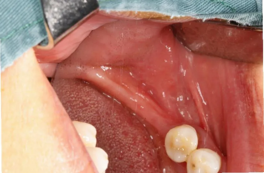
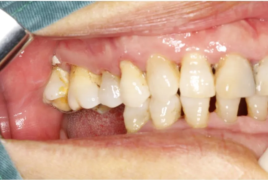
- Clinical Diagnosis: Mandibular dentition defect (46, 47)
Preoperative diagnosis of the patient – Intraoral photograph
Preoperative diagnosis of the patient – CBCT examination
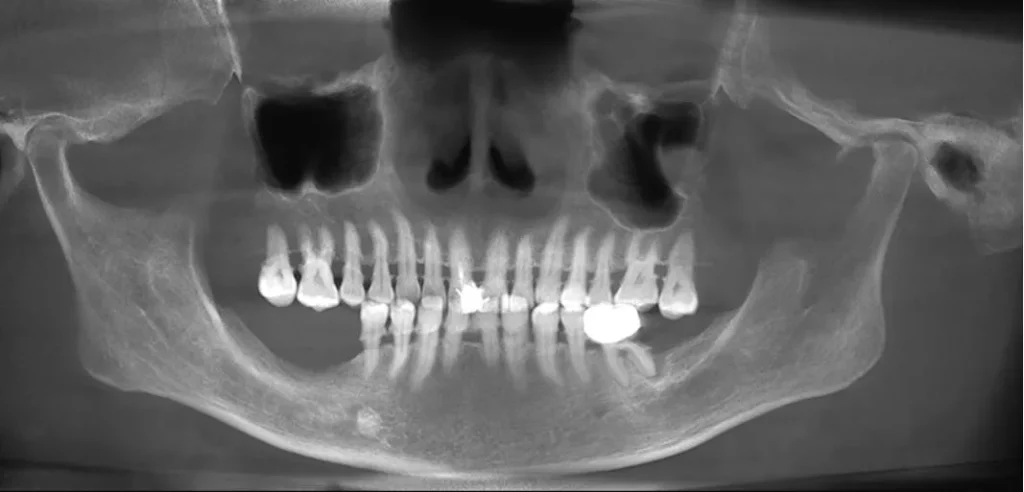
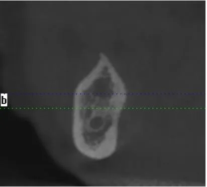
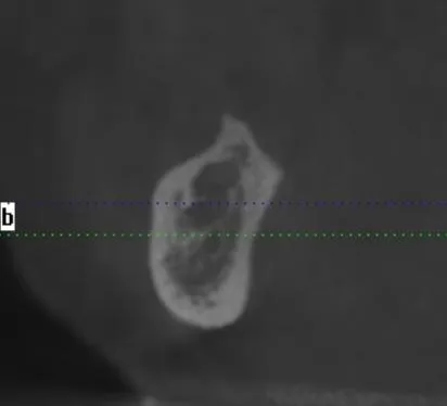
Panoramic CBCT reconstruction shows complete edentulism at #46 and #47 (right mandibular first & second molar). Severe alveolar ridge resorption is observed in the edentulous area; adjacent teeth #45 and #48 are preserved. The inferior alveolar nerve canal is close to the residual basal bone, insufficient native bone precludes immediate implant placement.
Treatment Plan
- Step 1:Stage 1 implant surgery with simultaneous GBR
- Step 2:Stage 2 implant surgery
- Step 3:Implant impression
- Step 4:Implant prosthesis placement.
Preoperative Preparation
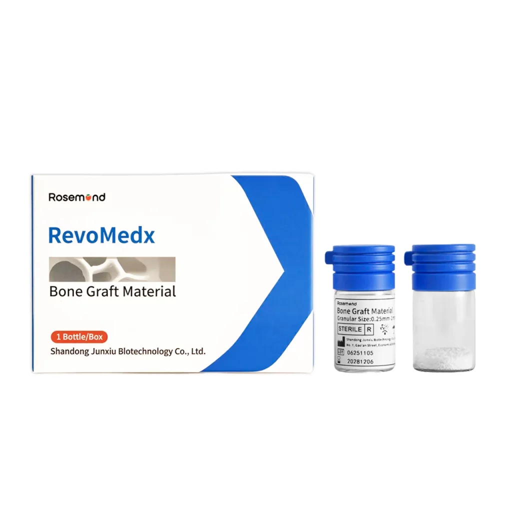
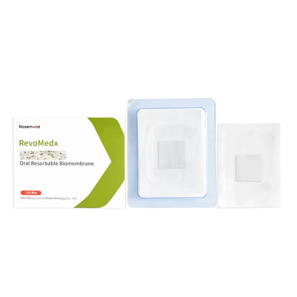
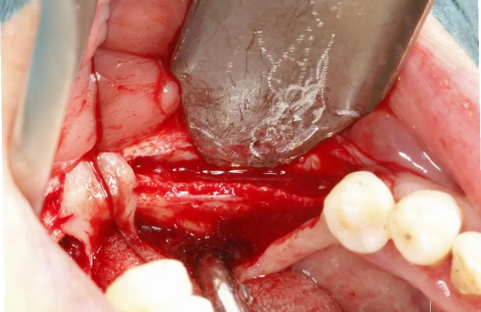
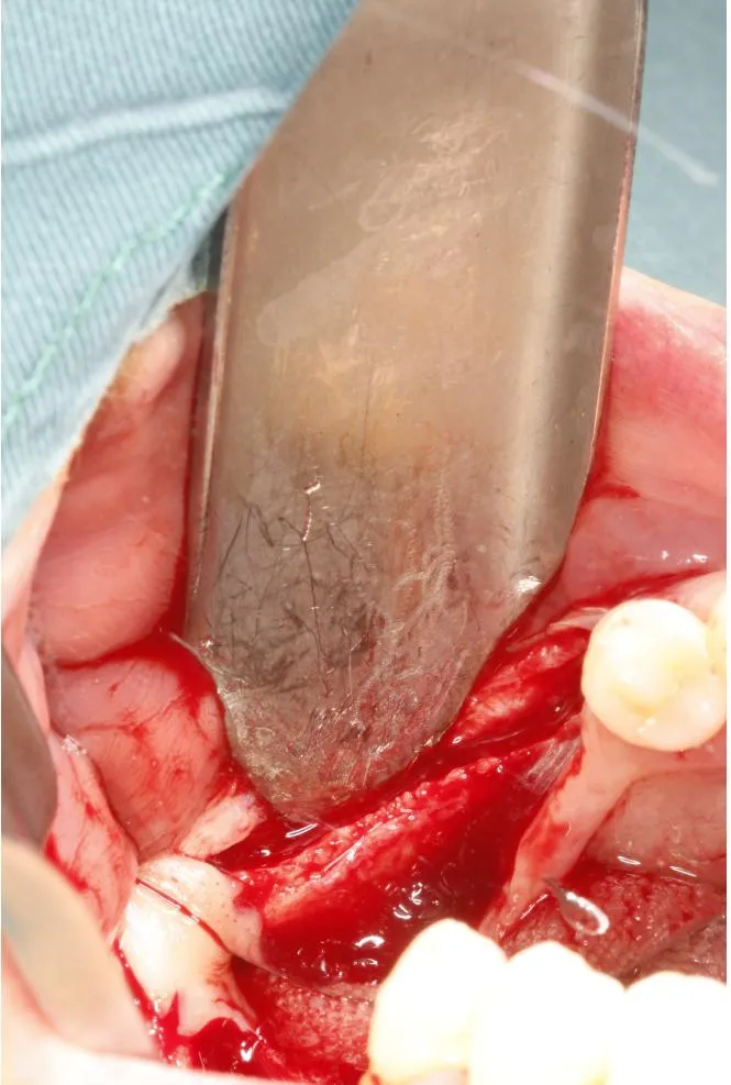
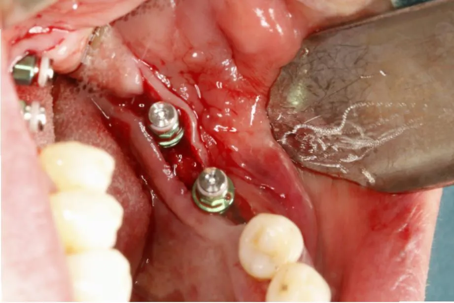
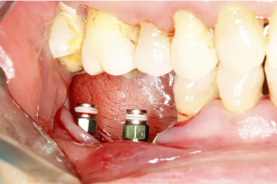
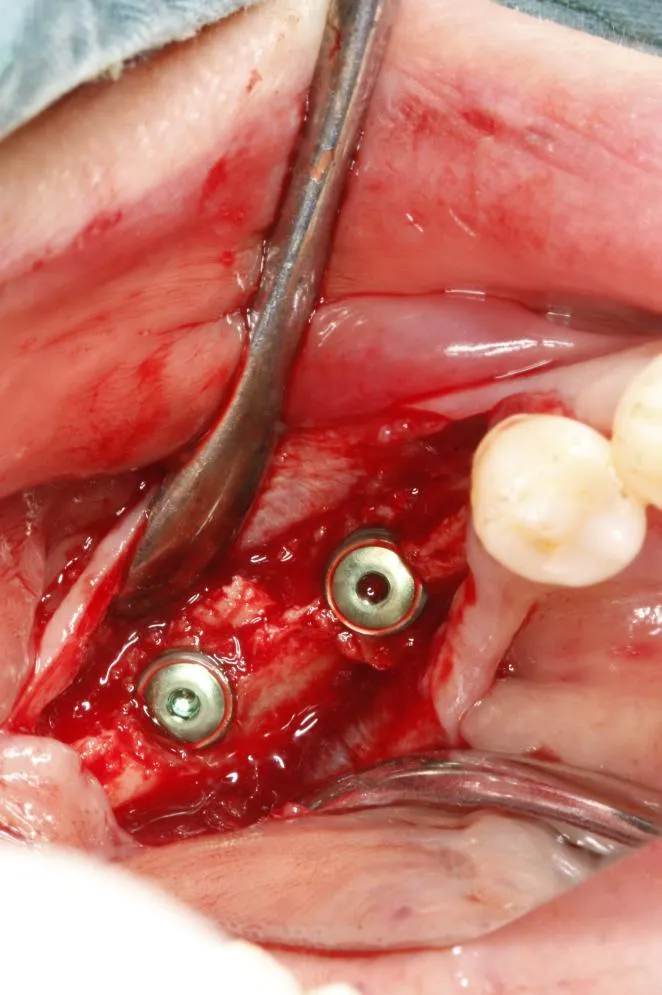
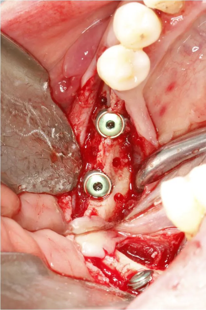
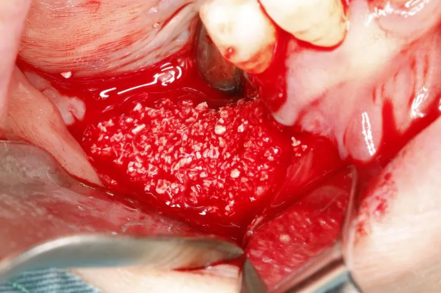
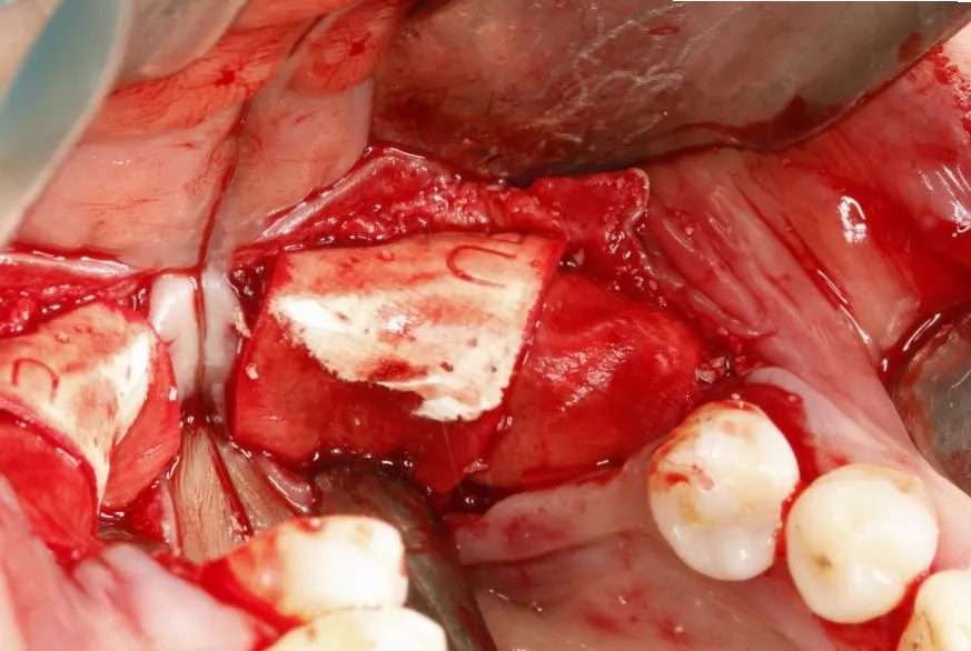
Intraoperative Photos – Teeth 46, 47
This set of photos documents the key steps of Guided Bone Regeneration (GBR) surgery with simultaneous implant placement at mandibular posterior sites (#46, #47):
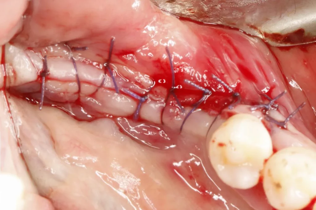
Postoperative Photos – Tooth positions 46, 47
Postoperative CBCT Examination – Tooth positions 46, 47
This is a postoperative CBCT image following Guided Bone Regeneration (GBR) surgery with simultaneous implant placement at mandibular posterior sites (#46, #47), used to evaluate bone augmentation outcomes and implant status:
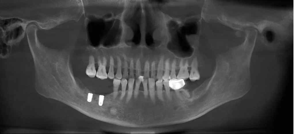
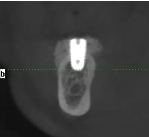
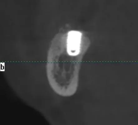
Two implants are clearly visible in the #46 and #47 edentulous sites, with precise positioning. The surrounding bone graft material shows homogeneous radiopacity and good integration with the host bone. No implant displacement, bone resorption, or inflammatory signs are observed, confirming stable surgical site anatomy.
Postoperative Photos At 6 Months – Tooth positions 46, 47
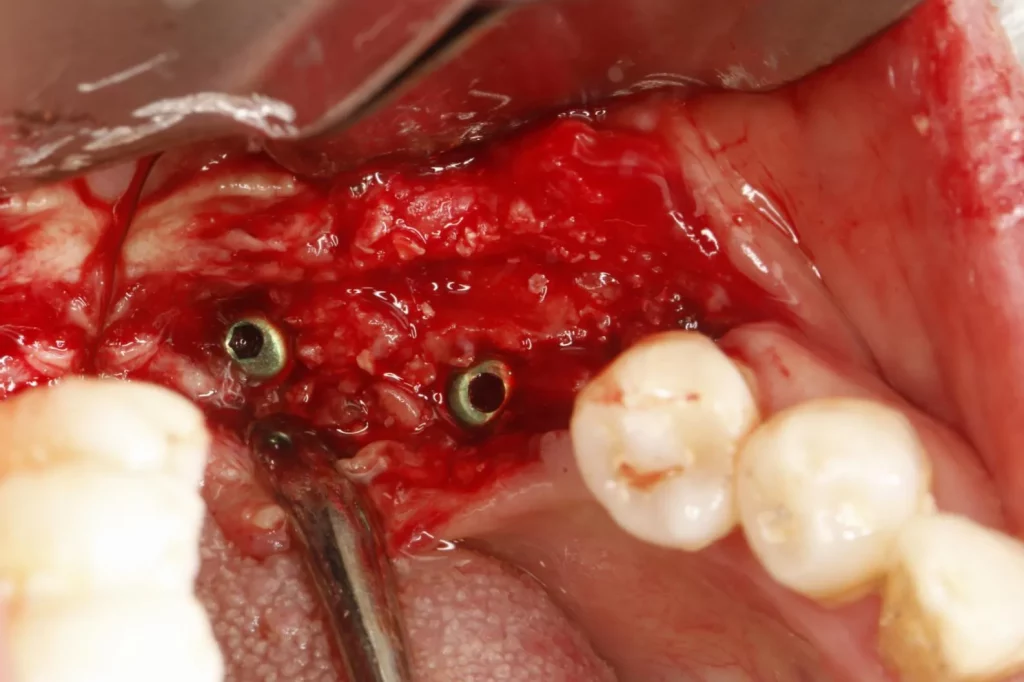
Postoperative CBCT Examination At 6 Months – Tooth positions 46, 47
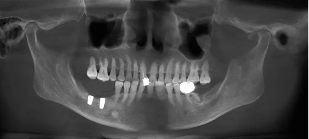
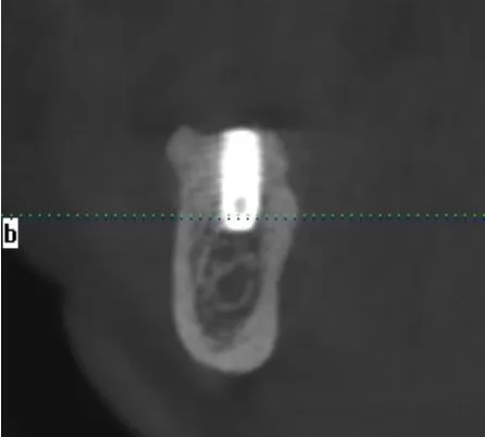
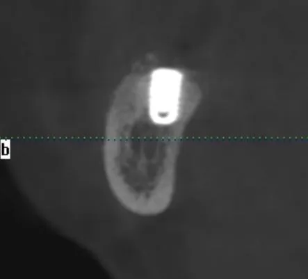
This is a post-restoration CBCT image after final prosthetic delivery, following Guided Bone Regeneration (GBR) surgery with simultaneous implant placement at mandibular posterior sites (#46, #47), fully validating the long-term outcomes of bone augmentation and implant rehabilitation.
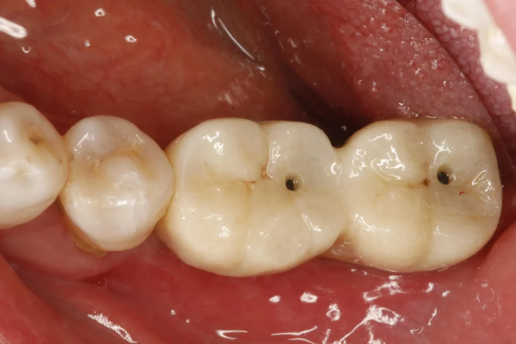
Post-insertion photos – Tooth positions 46, 47
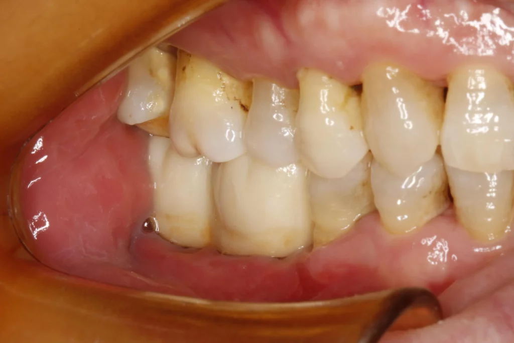
Post-insertion CBCT examination – Tooth positions 46, 47
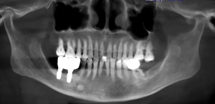
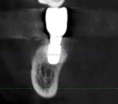
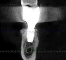
Summary
This final restoration image fully validates the excellent performance of Rosemond RevoMedx Natural Bovine Bone Graft Material+ Rosemond RevoMedx Oral Resorbable Biomembrane in GBR surgery:
The bone graft exhibits outstanding osteoconductivity and biocompatibility, effectively inducing new bone formation to provide sufficient osseointegration space and primary stability for implants.
The barrier membrane precisely prevents soft tissue ingrowth, maintaining a stable environment for bone regeneration and achieving durable reconstruction of bone defects.
The well-integrated bone-implant-prosthesis interface after final restoration provides strong evidence for the long-term success of implant rehabilitation in posterior mandibular bone defects.